


 CC BY
CC BY 29
29
УДК 616-006.85-07-089-053.2 Seung Hoon Choi
LONG-TERM FOLLOW-UP OF WILMS' TUMOR: A TWENTY-YEAR EXPERIENCE IN A SINGLE PEDIATRIC CENTER
Division of Pediatric Surgery, Department of Surgery Yonsei University College of Medicine (Seoul, Korea)
Keywords: Wilms' tumor, Demographics, Clinical, Threatment.
Wilms' tumor is a primary renal tumor of embryonic origin and usually presents in childhood. It makes up about 5—6% of all pediatric malignant neoplasms. Wilms' tumor is the fifth most common pediatric tumor, after CNS tumors, lymphoma, neuroblastoma, and soft tissue sarcomas. Unlike other malignant neoplasms, Wilms' tumors usually have a potential for cure. During the past twenty years, the treatment outcomes have improved, mostly due to the refinement of surgical technique and multimodal treatment regimen, including perioperative adjuvant chemotherapy and radiotherapy. We retrospectively studied pathologically confirmed Wilms' tumor patients at Yonsei University Medical Center Severance Hospital who underwent a nephrectomy during a 20-year period, from March 1986 to March 2005. Specific operations performed included ipsilateral nephrectomy and lymph node sampling for unilateral Wilms' tumor, and ipsilateral nephrectomy and contralateral partial ne-phrectomy or biopsy. We followed the NWTS (National Wilms' Tumor Study Group) protocol recommendation regarding chemotherapy, using EE-4A (pulse-intensive Vincristine + Actinomycin D for 18 weeks). If the tumor showed unfavorable histology, doxorubicin was added to the regimen (DD-4A, including doxorubicin for 24 weeks). If the tumor recurred after the initial course of chemotherapy, we adopted the ICE regimen (ifosfamide + carboplatin + VP-16). Postoperative radiation therapy was performed if the tumor was incompletely resected, or in the case of distant metastasis, tumor cell anaplasia, postoperative local relapse, the bilateral Wilms' tumor, or gross tumor cell emboli. We retrospectively analyzed the patients (based on admission records) in terms of their gender, age at operation, clinical signs and symptoms, associated anomalies, pathologic findings, clinical stage, postoperative morbidity and mortality, overall five-year survival rate, and disease-free survival rate. In addition, we also analyzed risk factors affecting tumor recurrence and patient survival. During the twenty-year period, 68 patients were pathologically diagnosed with Wilms' tumor after receiving a nephrectomy for a primary renal tumor. Those patients with clear cell sarcomas or rhab-doid tumors were excluded from this study. Mean age at diagnosis was 25 months (range: 2-130 months). Forty patients (58.8%) were male, and 28 (41.2%) were female. Male-to-female ratio was 1.4:1. Primary signs and symp-
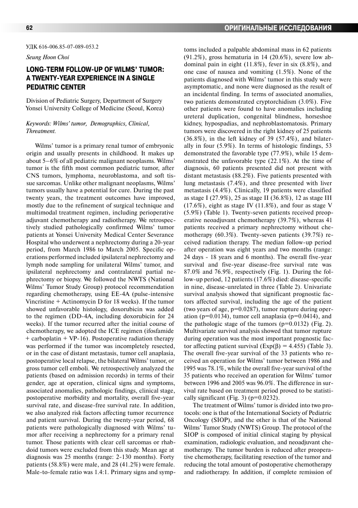
toms included a palpable abdominal mass in 62 patients (91.2%), gross hematuria in 14 (20.6%), severe low abdominal pain in eight (11.8%), fever in six (8.8%), and one case of nausea and vomiting (1.5%). None of the patients diagnosed with Wilms' tumor in this study were asymptomatic, and none were diagnosed as the result of an incidental finding. In terms of associated anomalies, two patients demonstrated cryptorchidism (3.0%). Five other patients were found to have anomalies including ureteral duplication, congenital blindness, horseshoe kidney, hypospadias, and nephroblastomatosis. Primary tumors were discovered in the right kidney of 25 patients (36.8%), in the left kidney of 39 (57.4%), and bilaterally in four (5.9%). In terms of histologic findings, 53 demonstrated the favorable type (77.9%), while 15 demonstrated the unfavorable type (22.1%). At the time of diagnosis, 60 patients presented did not present with distant metastasis (88.2%). Five patients presented with lung metastasis (7.4%), and three presented with liver metastasis (4.4%). Clinically, 19 patients were classified as stage I (27.9%), 25 as stage II (36.8%), 12 as stage III (17.6%), eight as stage IV (11.8%), and four as stage V (5.9%) (Table 1). Twenty-seven patients received preoperative neoadjuvant chemotherapy (39.7%), whereas 41 patients received a primary nephrectomy without chemotherapy (60.3%). Twenty-seven patients (39.7%) received radiation therapy. The median follow-up period after operation was eight years and two months (range: 24 days - 18 years and 6 months). The overall five-year survival and five-year disease-free survival rate was 87.0% and 76.9%, respectively (Fig. 1). During the follow-up period, 12 patients (17.6%) died: disease-specific in nine, disease-unrelated in three (Table 2). Univariate survival analysis showed that significant prognostic factors affected survival, including the age of the patient (two years of age, p=0.0287), tumor rupture during operation (p=0.0134), tumor cell anaplasia (p=0.0414), and the pathologic stage of the tumors (p=0.0132) (Fig. 2). Multivariate survival analysis showed that tumor rupture during operation was the most important prognostic factor affecting patient survival (Exp(P) = 4.455) (Table 3). The overall five-year survival of the 33 patients who received an operation for Wilms' tumor between 1986 and 1995 was 78.1%, while the overall five-year survival of the 35 patients who received an operation for Wilms' tumor between 1996 and 2005 was 96.0%. The difference in survival rate based on treatment period proved to be statistically significant (Fig. 3) (p=0.0232).
The treatment of Wilms' tumor is divided into two protocols: one is that of the International Society of Pediatric Oncology (SIOP), and the other is that of the National Wilms' Tumor Study (NWTS) Group. The protocol of the SIOP is composed of initial clinical staging by physical examination, radiologic evaluation, and neoadjuvant chemotherapy. The tumor burden is reduced after preopera-tive chemotherapy, facilitating resection of the tumor and reducing the total amount of postoperative chemotherapy and radiotherapy. In addition, if complete remission of
Table 1
Patient (n=68) Demographics and Clinical Parameters
Overal Survival of Wilms' tumor patients
Parameters Number
abs. %
Gender Male 40 58.8
Female 28 41.2
Age at diagnosis (years) <2 33 48.5
2-4 19 27.9
>4 16 23.5
Initial clinical presentation Palpable abdominal mass 62 91.2
Gross hematuria 14 20.6
Abdominal pain 8 11.8
Fever 6 8.8
Nausea/vomiting 1 1.5
Asymptomatic 0 0.0
Associated anomaly Cryptorchidism 2 3.0
Ureter duplication 1 1.5
Congenital blindness 1 1.5
Horseshoe Kidney 1 1.5
Hypospadia 1 1.5
Nephroblastomatosis 1 1.5
Tumor characteristics
Laterality Right 25 36.8
Left 39 57.4
Bilateral 4 5.9
Histology Favorable 53 77.9
Unfavorable 15 22.1
Initial Metastasis No 60 88.2
Lung 5 7.4
Liver 3 4.4
Clinical stage I 19 27.9
II 25 36.8
III 12 17.6
IV 8 11.8
V 4 5.9
1.00 .90 -.80 -.70 -.60 -.50 -
гч_
5 year OS = 87.0%
.40 x 0
_L
_L
_L
_L
48 96 144 192
Interval after operation (mon)
Recur Free Survival of Wilms' tumor patients
240
1.0 ~i
.9 -
.7 -.6 -.5 -
.4
5 year RFS = 76.9%
_L
_L
_L
_L
J
0
48
240
a metastatic lesion does occur after neoadjuvant chemotherapy, SIOP protocol can downstage the tumor, which may already present distant metastasis at initial diagnosis. Nevertheless, peritumoral adhesion secondary to necro-inflammatory changes after neoadjuvant chemotherapy makes precise dissection between tumor and normal tissue more difficult. However, it is that adhesion that also minimizes the likelihood of tumor rupture during operation and subsequent peritoneal tumor seeding and local recurrence. The SIOP protocol has a few pitfalls: neoadjuvant chemotherapy is performed without histopathologic confirmation, so there is a possibility that a patient with a benign or borderline tumor may receive chemotherapy. Likewise, patients with a more serious tumor (or tumors with highly malignant potential) may be undertreated, instead receiving a reduced dosage of chemotherapy. In the results of SIOP-9, 2% of the patients who received neoadjuvant chemotherapy without tumor biopsy did indeed have a benign tumor, and 3% were found to have a malignant renal
96 144 192
Interval after operation (mon)
Fig. 1. Overall survival (OS) and recurrence-free survival (RFS) of 68 Wilms' tumor patients (Overall five-year survival = 87.0%, five-year recurrence-free survival = 76.9%).
tumor other than a Wilms' tumor. Therefore, transcutaneous tumor biopsy is highly recommended in cases where preoperative neoadjuvant chemotherapy will be used as a treatment modality. The protocol for NWTS is composed of an initial nephrectomy and lymph node sampling, histological confirmation of the tumor, and pathologic staging followed by postoperative adjuvant chemotherapy. NWTS protocol has the advantage of proper chemotherapy relevant to stage of the tumor. However, if the tumor size is very large, or if the tumor has formed an adhesion to adjacent great vessels or other organs, resection is often impossible, and the chance of postoperative complications (such as intraoperative tumor rupture) increases. In this study, we didn't solely follow either the SIOP or NWTS protocol. Our treatments were divided into either initial nephrectomy or initial neoadjuvant chemotherapy. The latter therapy was adopted for stage IV patients who presented with either a distant metastatic lesion at the time of diagnosis, encasement of great vessels by the tumor, tight adhesion of the tumor to adjacent organs, or a large tumor not amenable to resection. For these tumors, we were able to perform a safer nephrectomy after initial neoad-juvant chemotherapy. One of the problems of the SIOP
Table 2
Mortality Cases After Surgery (n=12)
Sex Age (months) Histology Initial Meta. Pathologic Staging Late meta. Neoadj. CTx LR Cause of death Duration (months)
Disease specific death (n=9)
F 60 Anaplasia No II (sp.) No No Yes LR 22
F 120 FH No III(sp.) Liver No Yes LR, Liver 18
F 30 FH No II Liver Yes Yes LR, Liver 15
M 99 Anaplasia No III(sp.) Brain, Lung No Yes Brain, Lung 32
M 50 Anaplasia Liver IV Liver, testis No No Liver 28
M 102 FH No II (sp.) Lung Yes No Lung 63
M 7 UH No III Lung No No Lung 11
M 2 FH No III(sp.) No No Yes LR 54
M 78 FH Liver IV Lung No No Lung 10
Disease unrelated death (n=3)
M 49 UH Lung IV No Yes No D-CMP 28
M 31 FH No I No No No ALL (L-2) 162
M 5 FH No V No No No ARF 0.8
Abbreviations: Meta., Metastasis; Neoadj. CTx, Neoadjuvant chemotherapy; LR, Locoregional Recurrence; Sp., Tumor Spillage or Rupture During Surgery; FH/UH, Favorable Histology/Unfavorable Histology; D-CMP, Dilated Cardiomyopathy; ALL, Acute Lymphocytic Leukemia; ARF, Acute Renal Failure.
1 rvi
u
u
C
1 rvi
u
u
C
.7 .6 .5 .4
1.0 .9 .8 .7 .6 .5 .4
OS according to Patients' age
1 ............__
< 2 year
OS according to tumor spillage
> 2 year
1 rvi
u
u
C
_L
_L
p=0.0287 _I_
1.0
.9 -
.7 -
.5 -
_L
"4
Spill (-)
_L
Spill (+)
p=0.0134 _I_
_L
48 96 144 192
Interval after operation (mon)
OS according to Tumor cell anaplasia
240
Ri
Ana (—)
_L
_L
Ana (+)
p=0.0414
_L
1 rvi
u
u
C
1.0 i
.9 -
.7 -
.6 -
.5 -
_L
J
0
48
.4
48 96 144 192
Interval after operation (mon)
OS according to pathologic stage
240
1
I, II
_L
_L
III, IV
p=0.0132
_L
_L
J
240
96 144 192 240 0 48 96 144 192
Interval after operation (mon) Interval after operation (mon)
Fig. 2. Univariate analysis of prognostic factors affecting overall survival (OS) of Wilms' tumor patients (using log rank test) according to patients' age, tumor spillage, tumor cell anaplasia, and pathologic stage.
Table 3
Multivariate analysis of prognostic factors affecting overall survival of Wilms' tumor patients (Cox regression analysis)
Parameters B df. Sig. Exp (B) 95% CI for Exp (B)
Lower Upper
Age (>2 yr or <2 yr) 1.251 1 0.273 3.494 0.373 32.752
Tumor spillage1 1.494 1 0.031 4.455 1.145 17.341
Tumor anaplasia 1.412 1 0.070 4.104 0.891 18.905
Pathologic stage 0.630 1 0.091 1.877 0.905 3.892
Abbreviations: df., degree of freedom; Sig., significance; CI, Confidence Interval. 1 Tumor spillage was also the most important factor in recurrence-free survival.
Table 4
Clinical stage and pathologic stage of the 68 patients
Clinical Pathologic stage Total
Stage I II III IV V abs. %
I 13 6 0 0 0 19 27.9
II 9 7 9 0 0 25 36.8
III 1 7 4 0 0 12 17.6
IV 0 1 0 7 0 8 11.8
V 0 0 0 0 4 4 5.9
Total abs. 23 21 13 7 4 68 100.0
% 33.8 30.9 19.1 10.3 5.9
OS according to operation period
oi
1 'E
3
3
О
.7 -.6 -.5 -.4 -
1996-2005 (n=35) : 96.1%
1986-1995 (n=33) : 78.1%
_L
_L
p=0.0232
_I_
_L
Clinical stage is based on preoperative Ultrasonography (US), Computerized Tomography (CT), and physical examination Pathologic stage is based on tumor histopathology after operation.
protocol is overstaging. First, lymph node enlargement in radiologic study is often discovered intraoperatively to be secondary to nonspecific lymphadenopathy rather than metastasis of the tumor. Second, even if the tumor seems to be tightly adhered to an adjacent organ and determined unresectable upon radiologic evaluation, the tumor may in fact be easily separated from the adjacent organ, mainly because often the adhesion is actually a result of inflammation rather than tumor advancement. In this study, tumors were larger than those reported in other studies for a relative number of patients in clinical stages I and II. The median age at the time of operation was 25 months. These are due to earlier diagnosis of Wilms' tumor than in other studies. Thirty-three patients (48.5%) demonstrated a discrepancy between clinical stage and pathologic stage. These were divided into two groups: 18 patients had downstaging of tumor pathologic stage and 15 had upstaging of tumor pathologic stage (Table 4). The former condition was due to downstaging of the tumor after successful neoadjuvant chemotherapy or after a radiologically overstaged tumor was proven to be less advanced. The latter condition was due to over-staging of the tumor in the case of intraoperative tumor dissemination, or if the tumor cell showed unfavorable histology. In these circumstances, postoperative adjuvant therapy should be adopted as protocol dictates. In this study surgical rupture of the tumor was the most important prognostic factor affecting survival and recurrence (Relative Risk = 4.5).
0 48 96 144 192
Interval after operation (mon)
240
Fig. 3. Five-year Overall survival (OS) of the Wilms' tumor patients according to the operation period (using Log rank test p=0.0232). Five-year recurrence-free survival (not shown) also demonstrated a trend toward improvement (from 69.6% to 84.5%, p=0.1667).
Surgical rupture is defined as intraoperative tumor transsection and/or peritoneal dissemination of tumor cells during operation. However, it does not imply tumor dissemination after preoperative tumor biopsy or minimal tumor spillage. In general, tumor rupture seems to be highly related to local recurrence. In our multivariate survival analysis, tumor rupture was also the most important independent prognostic factor affecting local recurrence and survival of the patients. Considering tumor histology and pathologic stage, which were compatible with the results of NWTS-1, 2, 3 in which tumor rupture during operation is related to infra-diaphragmatic recurrence. If tumor rupture occurred during dissection, the tumor stage was upgraded, and postoperative chemotherapy or radiotherapy was performed according to the new pathologic staging.
There are several important surgical considerations:
1) A transverse incision should be made large enough to see the contralateral kidney with the transperitoneal approach being the preferred method (as opposed to the retroperitoneal approach); 2) Initial ligation of the proximal portion of renal vessels decreases blood loss during operation and minimizes the chance of hematolymphan-gitic metastasis; 3) Capsular tearing causes tumor cells to disseminate into the peritoneal cavity; and 4) Formal lymph node dissection of the perirenal, periaortic area
Table 5
Bilateral Wilms' tumor patients (n=4)
Sex Age (months) Preop bx. Neo RK LK Histology Post-op CTx Post-op RTx Recur Survival (months) RFS (months) Remark
F 38 + + Tot Par FH + + LK, Lung 142 28 —
F 40 + + Par Tot FH + + — 95 95 Horseshoe kidney
M 5 — — Par Tot FH — — — — — Death from ARF
F 10 + + Par1 Par FH + + - 5 5 -
Abbreviations: Pre-op bx., preoperative biopsy; Neo, Neoadjuvant Chemotherapy; RK, Right Kidney;
LK, Left Kidney; Post-op CTx, Postoperative Chemotherapy; Post-op. RTx, Postoperative Radiotherapy; RFS, Recurrence-free Survival; Tot, Total Nephrectomy; Par, Partial Nephrectomy; 1 Resection margin positive.
is unnecessary and even harmful (e.g. causing formation of chylous ascites), though it helps to select proper che-motherapeutic agents. In this study, the overall five-year survival was increased from 76% (between 1986 and 1995, n=33, early period) to 96% (between 1996 and 2005, n=35, later period). The pathologic stage and frequency of neoadjuvant chemotherapy were not statistically different in each period. This improved survival may introduce study bias, as the patients in the later period had a shorter follow-up period as compared with patients in the earlier period. On the other hand, this improvement in survival may be due to the fact that the number of patients who present with a tumor rupture during surgery has decreased among patients classified in a later stage (30.3% (10/33) vs. 11.4% (4/35), p=0.054).
There has been much controversy regarding the optimal treatment of bilateral Wilms' tumor. When planning treatment, the age of the patients and tumor histology should be primary considerations. Two important aspects to consider during surgery for the bilateral tumor are the complete resection of the tumor and the preservation of normal renal tissue and renal function during dissection (if possible) in order to prevent renal function deterioration. Until now, there has been no significant difference among survival rates of patients with a bilateral Wilms' tumor, patients who underwent an initial tumor biopsy and received neoadjuvant chemotherapy and surgery, and the patients who received an initial nephrectomy followed by postoperative adjuvant chemotherapy. We recommend an initial tumor biopsy with neoadjuvant chemotherapy, followed by surgery. After neoadjuvant chemotherapy, the tumor should be reevaluated for any changes in tumor diameter or changes in tumor advancement. If the size of the tumor remains unchanged, then surgery should be performed, i.e. a partial nephrectomy, including tumor or wedge resection.
Postoperative radiation therapy for the patients with a bilateral Wilms' tumor is selectively performed according to the effectiveness of previous therapy. In our study, four patients who were diagnosed with a bilateral Wilms' tumor (table 5) were treated with an initial unilateral nephrecto-my and contralateral partial nephrectomy or wedge biopsy, followed by postoperative adjuvant chemotherapy. Partial nephrectomies should be performed only if postoperative deterioration of renal function is unlikely. If postoperative renal failure dose occur, peritoneal dialysis and renal transplantation should be considered.
Recently, organ preservation surgery has been favored over aggressive surgery, even in the treatment of selective malignant neoplasms. The use of a partial nephrectomy in the treatment of Wilms' tumor has been a matter of debate. NWTS-5 does not recommend a partial nephrectomy, but patients with a unilateral, small-sized tumor diagnosed early may benefit from a partial nephrectomy, which is comparable to a conventional en-bloc resection of the tumor.
Benefits and toxicities of perioperative chemothera-peutic agents have long been debated, especially in cases involving young infants diagnosed before 12 months of age. The dosage should be reduced to half of the proper dosage. Dose reduction of chemotherapeutic agents in young infants not only reduced toxicity, but also maintained a treatment outcome comparable to conventional dosage. Regular liver function tests should be performed after chemotherapy treatment because veno-occlusive disease may occur following chemotherapy. Most of the patients with intestinal obstruction secondary to adhesion improve by non-operative management. One patient in our study received an operation for intestinal obstruction. This case comprises 1.5% of the total patients, and comprises 3.7% of patients who received adjuvant chemotherapy. This still represents a lower incidence rate than evident in other reports. Three patients in our study died after multimodal treatment secondary to causes other than cancer: one patient as a result of a dilated cardiomyopathy, one due to acute renal failure, and one due to acute lymphoblastic leukemia. Cardiomyopathy is related radiation therapy and/or doxorubicin toxicity. Acute renal failure is related to the abrupt reduction and deterioration of renal function after surgery. The incidence of a secondary malignancy increases during or after treatment, and is usually associated with the intensity of radiation therapy, doxorubicin use, and/or genetic predisposition.
Поступила в редакцию 25.10.05. 20-ЛЕТНИЙ ОПЫТ ИЗУЧЕНИЯ ОПУХОЛИ ВИЛЬМСА Seung Hoon Choi
Отделение детской хирургии университетского медицинского колледжа (Сеул, Корея)
Обобщен 20-летний опыт диагностики и лечения опухоли Вильмса. Наблюдали 68 больных, диагноз которым был поставлен в возрасте от 2 месяцев до 10 лет (40 мальчиков и 28 девочек). Отмечено повышение 5-летней выживаемости больных с 78,1% (1986-1995) до 96,1% (1996-2005). Обсуждается тактика предоперационного ведения пациентов и применение неоадъювантной терапии по протоколам SIOP и NWTS.
Pacific Medical Journal, 2005, No. 4, p. 62—66.
